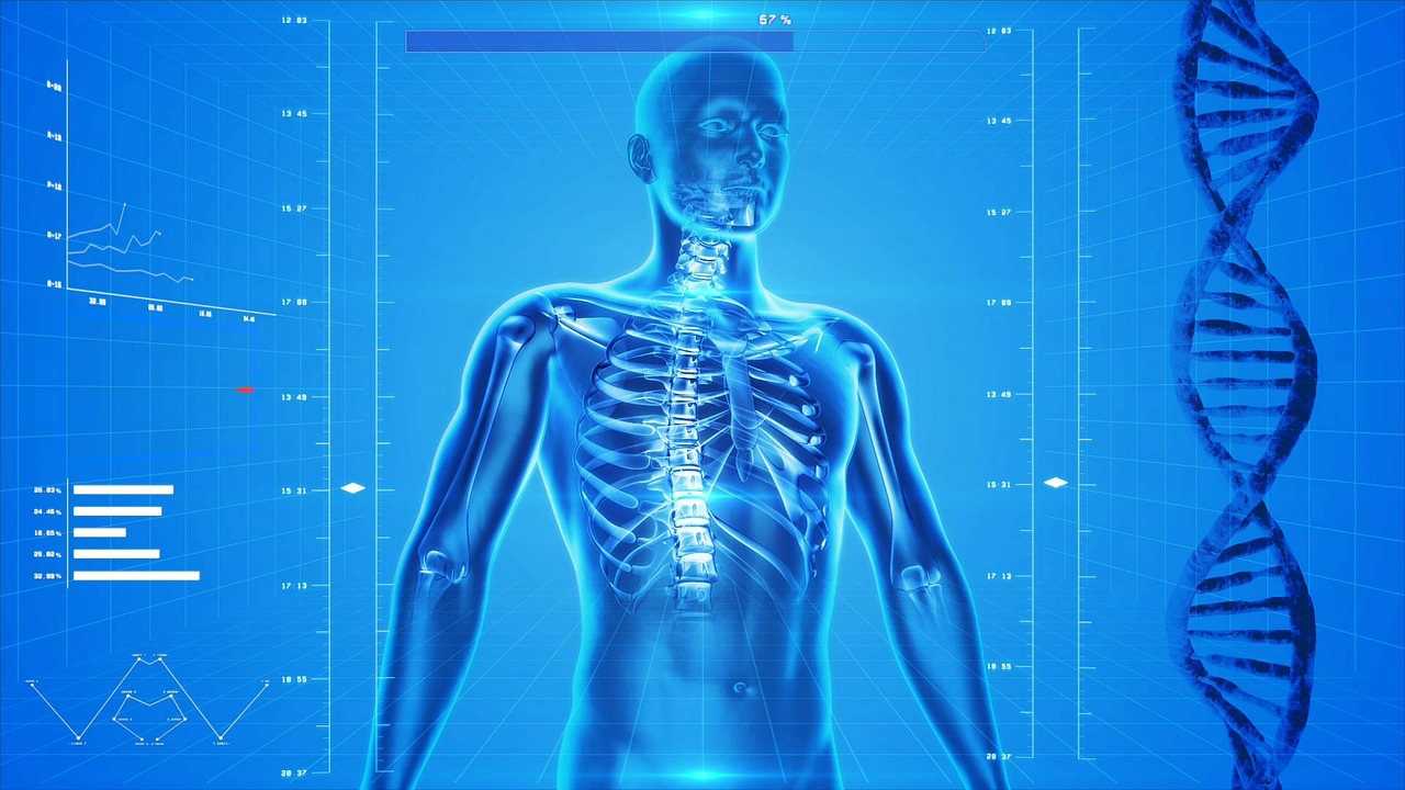
 As you are reading this, your skeleton is regenerating itself throughout every bone to replace old, micro-fractured bone with new bone formed in sheets with biomechanical properties similar to plywood.
This is achieved by specialised multi-nucleate osteoclasts (the only cells in the body that can dissolve hydroxyapatite crystals) using a proton pump similar to the stomach wall. These remove an area of damaged bone as they move across the surface of the honeycomb-like trabecular bone at the ends of long bones and in the vertebrae, or cut out a cylinder of bone in cortical bone.

Bone is replaced with layers of osteoid protein secreted by osteoblasts. Once outside the cell the osteoid is infiltrated with hydroxyapatite crystals consisting of calcium, phosphate and water molecules that provide bone rigidity.
This happens in all bones to regenerate them, a process taking five to 10 years making the skeleton five to 10 years’ old.
This regenerative mechanism does not maintain bone structure after the age of 30 because osteoclastic resorption occurs faster than osteoblastic bone formation due to osteoblast senescence, an active research area connected to other areas of cell senescence research.
We have pharmaceuticals available that, in conjunction with lifestyle modification, not only stop progression but can result in skeletal regeneration.
The MBS bone density schedule (developed by a committee I chaired back in 1995) lists the conditions predisposing to accelerated bone loss. Age-related bone senescence, exacerbated by ovarian or testicular failure, is the most common.
The question is not whether it can be done, but whether it is worth the time and cost to reduce fracture risk. The best way to decide this is by a dual energy bone structure examination (DXA) that, with extra demographic data, provides a reasonably accurate fracture risk over five years. Many use a 10% risk to advise the addition of pharmaceuticals to lifestyle advice.
Ideally your bone density provider will provide an easily readable image to correctly identify reference area of spine, hip or forearm and will provide a graph of where the patient is in relation to the normative data. These graphs act as an important explanatory aid to improve patient compliance and improve risk identification.
The usual pharmacological antiosteoclast medication must include calcium and vitamin D. More severe skeletal damage may benefit from osteoblast stimulatory medication in addition to certain antiosteoclast medications.
Osteoporosis is an important disease of older patients due to osteoblast senescence as well as younger individuals with disease predisposing to rapid increased bone turnover and associated relative osteoblast senescence. In all categories, it is possible to stop bone loss and, if necessary, regenerate the skeleton by using the inherent processes developed over our biological evolutionary history.
Key messages
- The skeleton can regenerate itself but loss exceeds this ability after age 30
- Osteoporosis is a treatable condition
- Bone scanning informs treatment decisions
References available on request.
Questions? Contact the editor.
Author competing interests: nil relevant disclosures.
Disclaimer: Please note, this website is not a substitute for independent professional advice. Nothing contained in this website is intended to be used as medical advice and it is not intended to be used to diagnose, treat, cure or prevent any disease, nor should it be used for therapeutic purposes or as a substitute for your own health professional’s advice. Opinions expressed at this website do not necessarily reflect those of Medical Forum magazine. Medical Forum makes no warranties about any of the content of this website, nor any representations or undertakings about any content of any other website referred to, or accessible, through this website.

