

 A 48-year-old man presented to our eye clinic with painless decline in his vision over the past 3 weeks. He has a history of advanced stage B-cell lymphoma.
Initial Presentation: A man with lymphoma and white eyes.
Description: A 48-year-old man came to our eye clinic because his vision had been deteriorating over the previous 3 weeks; he had no pain and no other eye symptoms.
Need more information?
In his medical history, he had been diagnosed with advanced stage, aggressive B-cell lymphoma—with no indication of CNS involvement: his cerebrospinal fluid had shown no cancer cells. He had been treated with systemic chemotherapy, with rituximab and intrathecal methotrexate prophylaxis for six cycles over a 110-day period, resulting in a complete remission; his last dose of his treatment was given 88 days before the onset of his vision problems.
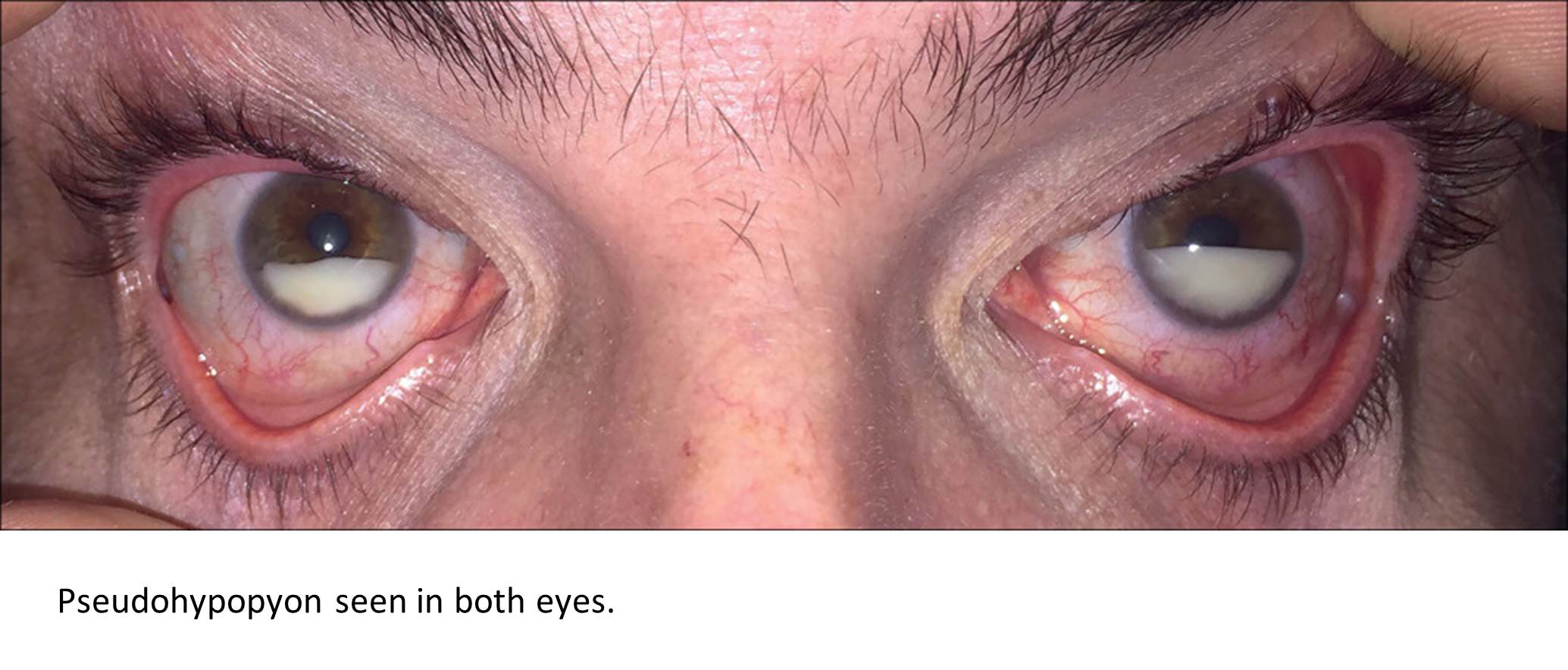
On examination, we found his visual acuity was 20/20 in the right eye and 20/60 in the left. Intraocular pressures were elevated in both eyes—43 mm Hg in the right and 52 mm Hg in the left (normal range 10–21). Both eyes showed mild conjunctival injection and corneal oedema, and the anterior chamber of both eyes contained a high concentration of cells—grade 4, severe cellularity—a layered pseudohypopyon, but no signs of flare or protein transudate (figure). The vitreous humour showed no cellularity or haze, and the patient’s retina and choroid were normal.
What is the most likely diagnosis?
- Bilateral eye infections
- Ocular involvement by systemic lymphoma
- Non-neoplastic uveitis
- Recent ocular steroid injections
 Answer: Ocular involvement by systemic lymphoma
Breakdown: We were concerned that he might have ocular metastasis—given the recent history of B-cell lymphoma—and a sample of aqueous humour confirmed our suspicions (appendix). The patient was again treated with rituximab and high-dose methotrexate. At follow-up, 2 months later, his vision was 20/20 in the right eye and 20/30 in the left. The pseudohypopyon had completely resolved in the right eye and was much reduced in the left; his intraocular pressures returned to within normal range using topical therapy (appendix). Neuroimaging and CSF studies remained unremarkable throughout the course of treatment. However, the lymphoma recurred—involving the CNS—and 15 months after the onset of his visual symptoms, he died.
A hypopyon—also known as sterile pus—is produced because of toxins, which are usually produced by pathogens, causing the collection of leucocytes in the anterior chamber of the eye. It can occur with a corneal ulcer, Behçet’s disease, endophthalmitis, and as an adverse reaction to some drugs—including rifabutin.
Pseudohypopyons can be caused by metastatic cells: they have been reported with leukaemias, lymphomas, and retinoblastomas. Patients with pseudohypopyons—unlike eye infections—typically have no pain or redness. Metastatic diseases in the eye can involve infiltration of the aqueous or vitreous humour, retina, or choroid. Mechanical blocking of the aqueous humour outflow by metastatic cells can result in increased intraocular pressures—as occurred in our patient. As is well known, but cannot be repeated often enough, eye problems should prompt a thorough history and an investigation for a primary cancer or for referral for further treatment in a patient with a known malignancy.
Source: All content for this “Diagnostic Puzzle” was sourced from The Lancet. The article was written by Siva S Radhakrishnan Iyer, MD, Zhongbo Jin, MD, Robert P Seifert, and MD, Prof Nam H Dang, PhD.
Original article is available at:
https://www.thelancet.com/journals/lancet/article/PIIS0140-6736(20)30610-3/fulltext
For more Diagnostic puzzles, go to: Picture Quiz Gallery (thelancet.com)