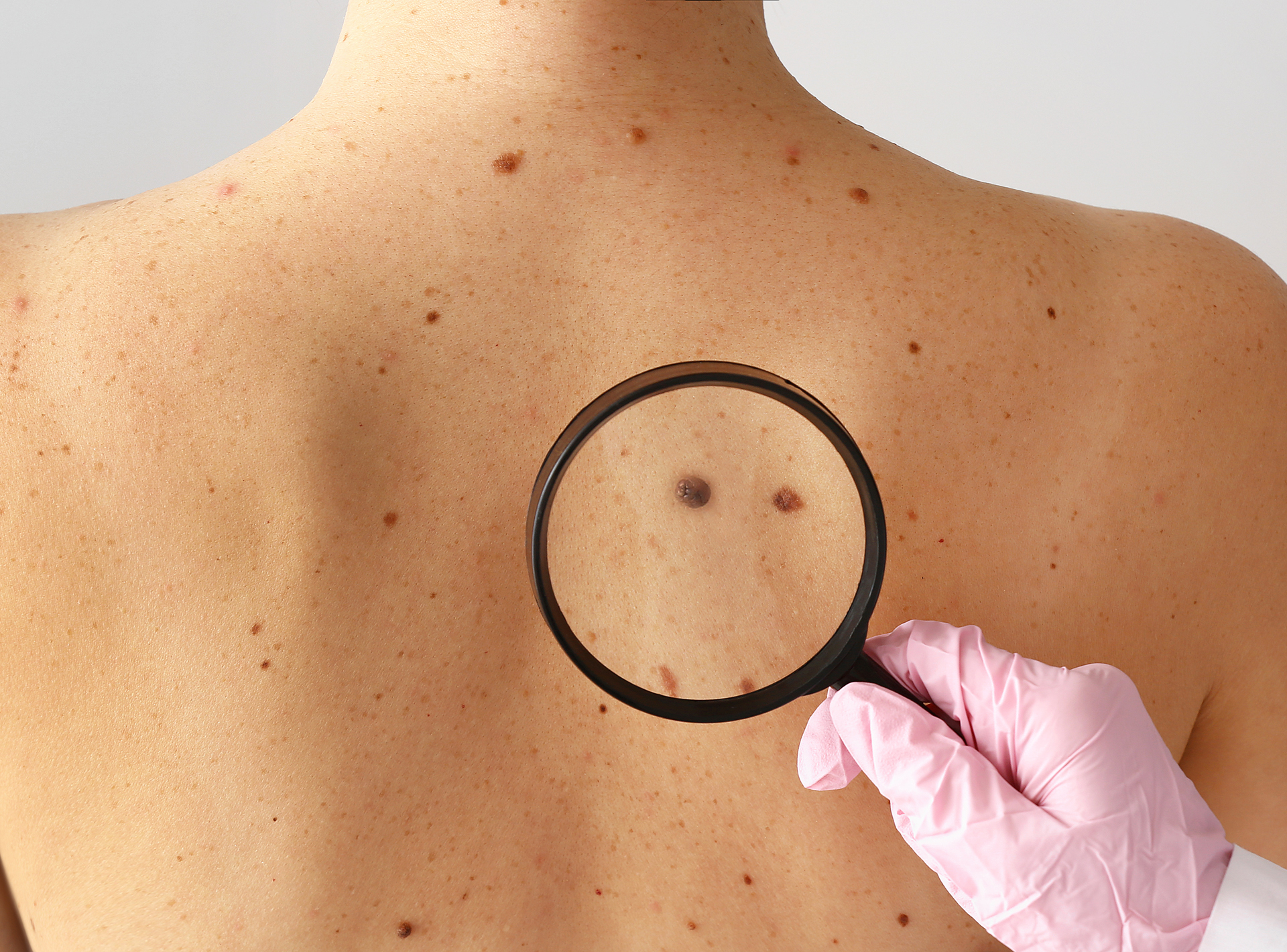
 An expert panel of internationally recognized dermatopathologists have developed a new classification system for melanoma.
According to the study, published January 2023 in JAMA, the Melanocytic Pathology Assessment Tool and Hierarchy for Diagnosis schema (MPATH-Dx), makes skin biopsy pathology reports easier for patients and medical teams to understand.
With an estimated 17,756 Australians diagnosed with melanoma in 2022 – or 11% of all new cancer cases and the third most detected cancer that year – a simple, standardized classification system for melanocytic lesions is increasingly important to aid pathologists and clinicians, who examine millions of tissue-samples each year.
Lead author Dr Raymond Barnhill from the Curie Institute and UFR of Medicine at the University of Paris, said that the team examined and made improvements to the original MPATH-Dx system (developed back in 2014) by incorporating feedback from U.S. dermatopathologists participating in an NIH-funded study and from members of the International Melanoma Pathology Study Group.
“Skin biopsies, particularly those suspicious for melanoma, are some of the most complicated for pathologists to diagnose,” Dr Barnhill said.
“Furthermore, pathology reports for skin biopsies are often filled with varied and complicated medical terms that can make a diagnosis or treatment plan unclear for medical providers, and with increased access to medical records and other digital resources, patients now share in the confusion and growing unease when reading their own skin biopsy reports.”
Although histopathology has functioned as the gold standard for diagnosis of cutaneous melanocytic lesions for decades, many reports over the years have called attention to a striking discordance in the interpretation of some lesions.
“A 2017 study by Elmore et al, to our knowledge the largest and most comprehensive of its kind, has confirmed that histopathological diagnosis across the spectrum of atypical and dysplastic nevi, including thin melanoma, is neither accurate nor reproducible, and these findings have significant implications for patient care,” Dr Barnhill explained.
“However, it is important to emphasize that a major factor accounting for such poor diagnostic concordance is the lack of established, agreed on, objective, and reproducible histopathological criteria along this continuum of lesions.
“Until more objective histopathologic breakpoints are delineated by precise correlation with genetic alterations and patient outcomes, diagnostic agreement will remain suboptimal.”
Even though the original MPATH-Dx made provisions for other types of melanocytic lesions, increasing knowledge in recent years about other pathways to melanoma and the appearance of the 4th edition of the WHO Classification of Skin Tumours highlighted the need to modify the original schema.
The updated schema, MPATH-Dx Version 2.0, simplifies the diagnoses of all melanocytic skin lesions undergoing biopsy into four classes, ranging from benign, minimal risk lesions, such as banal nevi in Class 1, through to the much higher risk Class IV, which includes thick, invasive melanoma cases that require appropriate surgical removal and additional evaluation for metastasis.
Co-author Dr Joann Elmore, professor of medicine at the David Geffen School of Medicine at UCLA, and member of the UCLA Jonsson Comprehensive Cancer Centre, explained that the MPATH-Dx schema also suggested guidance on how to best treat the patient for each of the four classes.
“Pathology reports can be confusing for both patients and the primary care physicians and this new tool will help provide clarity in what the pathology diagnosis is to these end users, along with simple guidance on the risk level of the lesion and appropriate treatment,” Dr Elmore said.
“In the past, primary care physicians might biopsy a patient’s suspicious looking skin lesion and receive a pathology report that states: ‘junctional dysplastic nevus with low-grade atypia’ and the primary care physician might not know what this means for the patient regarding underlying risk of melanoma and what, if any, additional treatment, or surveillance, is best for this patient.
“But when pathologists’ use the MPATH-Dx schema as an adjunct tool, the pathology report would include additional summary information stating ‘MPATH-Dx Class I, no further treatment required,’ thus reducing provider confusion and patient anxiety.”
The new version also has clearly defined histopathological criteria for classification of classes I and II lesions; specific provisions for the most frequently encountered low–cumulative sun damage pathway of melanoma progression, as well as other, less common pathways to melanoma; provides guidance for classifying intermediate class II tumours vs melanoma; and recognizes a subset of pT1a melanomas with very low risk and possible eventual reclassification as neoplasms lacking criteria for melanoma.
“Importantly, this schema is meant to be a flexible adjunct to existing nomenclatures or classification systems for benign melanocytic lesions, not a replacement,” Dr Barnhill said.
“For example, a pathologist may continue to use his or her own terminology and protocol for the grading of atypical nevi and then place (or map) the individual lesion into the appropriate MPATH-Dx class I or class II based on guidelines and the need for re-excision or not.
“We expect that the implementation of the new revised MPATH-Dx V2.0 schema into routine practice will provide a robust tool and adjunct for standardized diagnostic reporting of melanocytic lesions and management of patients to the benefit of both health care practitioners and patients.”

