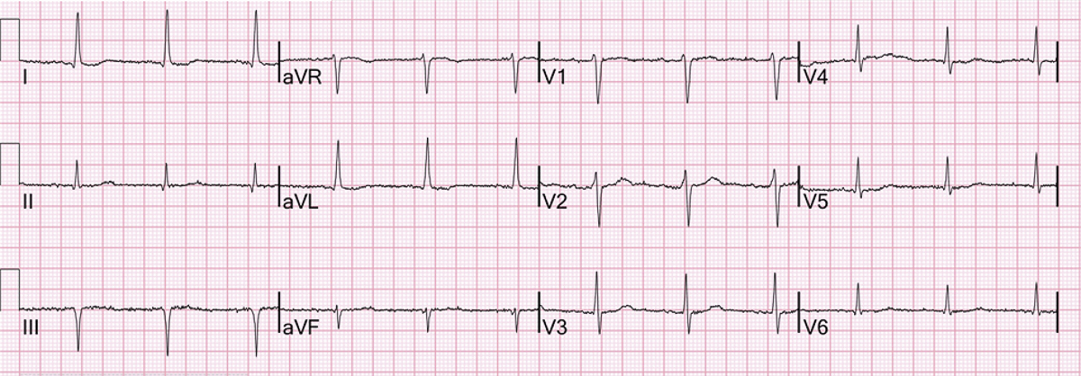
Cardiologists are regularly referred patients based on incorrect ECG machine reports, with misdiagnosis of the heart rhythm or overdiagnosis of prior myocardial infarction being the most common. The referring doctor may not have tried to interpret the ECG or be concerned about the medical or medicolegal consequences of disagreeing with the machine.

Often, faxing or emailing a high-quality copy of the trace to a cardiologist will resolve the issue immediately (a photo sent from a phone can suffice but is not as good), though not every cardiologist is a super whiz on ECG interpretation! Machine accuracy is improving, but algorithms vary between machines. No studies compare devices, primarily because manufacturers are reluctant to expose their patented algorithms.
Arrhythmias, pacemaker rhythms
ECG machines have about a 95% positive predictive accuracy for identifying sinus rhythm, falling to 53.5% for non-sinus rhythm. The problem in both situations relates to difficulties recognising small amplitude P waves, or P waves hidden by noise, QRS complexes or T or U waves.
Over-interpretation of atrial fibrillation is common, most usually due to frequent atrial ectopics (see figure). Overdiagnosis can result in administration of potentially harmful treatment.
Pacemaker rhythms are very poorly recognised, being interpreted as myocardial infarction, left ventricular hypertrophy or conduction disturbances mostly left bundle branch block.
IHD, LVH and prolonged QT
When an ECG is done in the setting of chest pain, wide variations exist in both over-call of ST elevation and under-diagnosis. ST elevation due to early repolarisation can be either a relatively benign normal variant or in rare cases a marker for risk of sudden death.
Pericarditis causes ST changes associated with chest pain. Very early in the course of myocardial infarction, the ECG can look completely normal, and acute changes can be masked by conduction abnormalities, particularly left bundle branch block.
History and the clinical context are more reliable when managing a patient presenting with chest pain. Act with caution, and do not delay treatment by measuring the troponin level before referring the patient to ED.
Old inferior wall myocardial infarction is a common ECG machine misdiagnosis, usually based on Q waves detected in leads III and aVF. This often results from a more horizontal heart position because of body habitus, with the Qs disappearing with the ECG recorded during held deep inspiration, changing the position of the heart relative to the leads.
If there is no significant Q in II, prior inferior wall infarction is unlikely. Poor anterior lead R wave progression is commonly interpreted as “possible anteroseptal myocardial infarction” but is usually an overdiagnosis. If the clinical context suggests prior MI as a possibility, then echocardiography is the best way of ruling this in or out.
Diagnosis of left ventricular hypertrophy is poor both by man and machine. Echocardiography is a much better tool. Accurate measurement of the QTc interval is difficult even for experts and the clinical context is everything.
If your machine measures the QT interval as prolonged, first check electrolytes and for any medications (e.g. psychotropic drugs, erythromycin or ketoconazole) which may be contributing. Remember that medication might be unmasking a genetic abnormality. If the patient has syncope, or a family history of syncope or sudden death, refer significant QT prolongation to a cardiac electrophysiologist.
Key messages
- Don’t blindly trust the ECG machine, make your own interpretation
- Clinical ECG Interpretation by Dr Araz Rawshani is a good online tool: https://ecgwaves.com/
- Get cardiologist input if unsure
References available on request.
Questions? Contact the editor.
Author competing interests: nil
Disclaimer: Please note, this website is not a substitute for independent professional advice. Nothing contained in this website is intended to be used as medical advice and it is not intended to be used to diagnose, treat, cure or prevent any disease, nor should it be used for therapeutic purposes or as a substitute for your own health professional’s advice. Opinions expressed at this website do not necessarily reflect those of Medical Forum magazine. Medical Forum makes no warranties about any of the content of this website, nor any representations or undertakings about any content of any other website referred to, or accessible, through this website.

