ED: Does the new NIPT replace the 12-week ultrasound scan?” The answer is ‘no’. We ask pathologist Dr Narelle Hadlow and obstetric radiologist Dr Anjana Thottungal to explain why – to read Narelle’s views click here: https://mforum.com.au/non-invasive-prenatal-testing-nipt/ . Here are Anjana’s views.
Cell free DNA testing cannot rule out structural abnormalities that occur during the development of a fetus, which is the most common cause of congenital disorders.
Since the introduction of non-invasive prenatal testing (NIPT), an increasing number of women are missing out on detailed 12 weeks scan, due to a misunderstanding by patients and perhaps inadequate information by health professionals during counselling early in pregnancy. This trend could potentially increase the number of pregnancies where the initial diagnoses of major abnormalities are being made at 18-20 weeks anatomy scan.
Important considerations
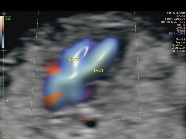
Major technological advances over the past 20 years in the detailed scan at 12-14 weeks (first trimester scan), have improved identification of chromosomal abnormalities and structural malformations of the fetus.
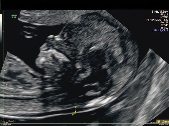
First trimester nuchal translucency (NT) measurement (See graphics) has evolved beyond its role for chromosomal anomaly screening.
Detailed imaging of a fetus less than 16 weeks old is now possible with the advent of high-resolution transabdominal and transvaginal ultrasound probes.
The threshold is lower for using transvaginal scans that provide detailed and high resolution images when the fetus is in a difficult position, in women with higher body mass index and in women at high risk for congenital malformations. (First trimester scans also have a role in the early prediction and risk assessment of early onset preeclampsia and preterm labour.)


The advantages of a fetal anatomy survey at 12-14 weeks
- Ability to see the complete fetus in one view.
- Increased fetal mobility allows imaging from different angles.
- There is a lack of boney ossification obstructing views.
- High-resolution transvaginal ultrasound brings the transducer closer to fetal organs.
- The number of soft markers and false positives is much lower at the early scan, limiting parental anxiety.
- Severe anomalies are detected early when the mother has not yet felt movements so that termination involves less psychological trauma for parents. There is also less risk for the mother.
What are the challenges of 12-14 week scan?
- Small size of fetal organs.
- Need to combine the abdominal and transvaginal approaches in some patients.
A recent study covered the effectiveness of the 12-14 week scan for early diagnosis of fetal congenital anomalies. It concluded that an early scan performed by a competent sonographer can detect about 45% of the prenatally diagnosed anomalies, including all lethal ones and 100% of those expected to be detected at this gestation (1).

It concluded that early detection of congenital anomalies was important and also confirmed the value of increased nuchal translucency (NT) in over 50% of the early-diagnosed anomalies, as a marker of abnormal development – beyond the role of NT in screening for chromosomal anomalies.

Key Messages
- Cell free DNA testing has brought greater focus on the value of the first trimester 12-14 week scan.
- NIPT and the first trimester scan, are principally focused on chromosome disorders and structural malformations, respectively.
- To effectively use these screening methods, both expectant mothers and their health professionals need to be aware how NIPT and 12-14 weeks scan complement each other.
References:
- Effectiveness of 12-13 weeks scan for early diagnosis of fetal congenital anomalies in the cell free DNA era. MJA Kenkhuis, M Barker, F Bardi, F Fontanella et al, Ultrasound Obstet Gynecol 2018; 51: 463-469
Further reading
First Trimester Ultrasound diagnosis of fetal abnormalities / Alfred Abuhamad, Rabih Chaoui 1st edition 2018
Disclaimer: Please note, this website is not a substitute for independent professional advice. Nothing contained in this website is intended to be used as medical advice and it is not intended to be used to diagnose, treat, cure or prevent any disease, nor should it be used for therapeutic purposes or as a substitute for your own health professional’s advice. Opinions expressed at this website do not necessarily reflect those of Medical Forum magazine. Medical Forum makes no warranties about any of the content of this website, nor any representations or undertakings about any content of any other website referred to, or accessible, through this website.

